Capillary Microscope
Eleftheria Foundation & Dr. Stephen Quake
Overview
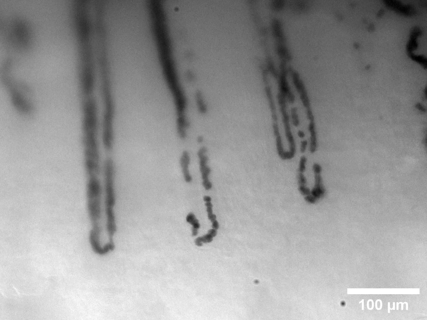
This project focuses on designing a microscope to image white blood cells flowing through nailfold capillaries — the tiny blood vessels visible at the base of the fingernail. By watching individual blood cells move through these capillaries, we can study microvascular health non-invasively.
The Finger-Lock
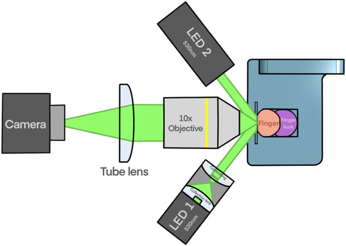
A key challenge in capillary microscopy is motion: even small finger movements blur the video. I invented an inflatable "finger-lock" that stabilizes the finger under the microscope while also applying controlled pressure to modulate blood flow velocities. This device enables both clearer imaging and the study of how capillary flow responds to pressure changes.
Data Collection
I wrote and received approval for an IRB protocol to study blood flow in healthy individuals and those with vascular conditions. Over the course of the project, I recruited and tested 82 participants.
Data & Analysis
Analysis Pipeline
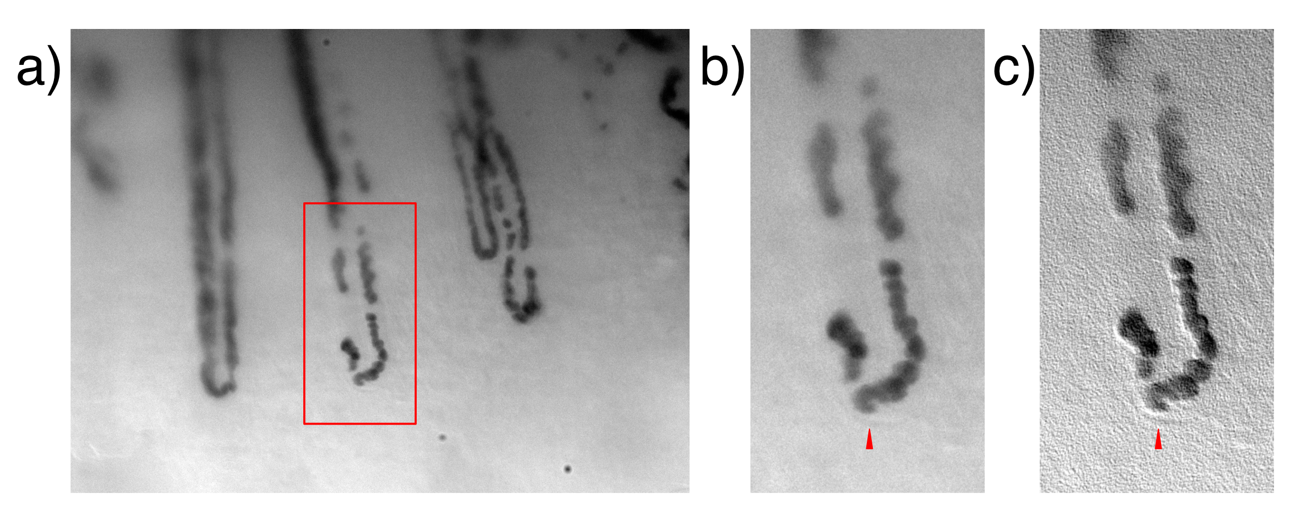
I developed an open-source Python and ImageJ pipeline (capillary-flow) to stabilize video, segment capillaries, and extract blood flow velocities from each frame. The pipeline handles the full workflow from raw video to quantitative flow measurements.
Publication
Forst ML, Rincon G, Levy JH, Cornfield DN, and Quake SR. An inflatable "finger-lock" for stabilizing nailfold capillary videos and modulating blood flow velocities. Review of Scientific Instruments. 2025; 96(8):083702.